Wellaholic Research: Microneedling for the Treatment of Scars
Table of Contents
TL;DR Summary
- What is Microneedling? – Tiny needles create micro-injuries to stimulate collagen production.
- Scar Reduction Benefits – Improves skin texture, reduces scars, and boosts collagen.
- Suitable for Various Scars – Effective for acne, surgical, and traumatic scars.
- Procedure Effectiveness – Results in smoother, firmer, and rejuvenated skin.
- Minimal Downtime – Quick recovery with minimal discomfort after treatment.
- Professional Microneedling – Best results are achieved through professional dermatological treatments.
Background
Microneedling is a beacon of hope for many people dealing with scars in the aesthetic world. At Wellaholic, we’ve not only applied this treatment but also dedicated time to understanding its effects deeply.
Microneedling, with its simple premise of using fine needles to create tiny punctures in the skin, surprisingly fosters natural healing. It’s this process that can dramatically improve the appearance of scars.
After conducting thorough research at Wellaholic, I have uncovered a wealth of convincing evidence that supports its numerous benefits.
In this article, I will discuss our findings about how microneedling can help reduce the visibility of scars.
Introduction
Microneedling is a used in dermatology for skin rejuvenation, tightening, scar treatment, and hair growth. Microneedling is often chosen over laser procedures for people with darker skin tones (Fitzpatrick IV to VI) because it causes less post-inflammatory hyperpigmentation.
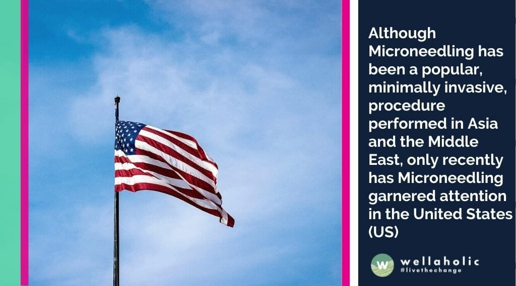
Although Microneedling has been a popular, minimally invasive, procedure performed in Asia and the Middle East, only recently has Microneedling garnered attention in the United States (US). Since the first clinical descriptions of subcision and “needle dermabrasion” (using a tattoo gun without ink), the production of Microneedling devices has flourished.1,2 In the US, Microneedling devices exist as both rollers, stampers, and pens (electrically powered or otherwise), and can be combined with radio frequency (RF) in an effort to deliver energy below the epidermal surface – known as fractional radio frequency microneedling (FRF-MN), thus avoiding epidermal damage and subsequent dyspigmentation.3
Microneedling devices vary based on their needle length (i.e. depth of skin penetration), diameter, density and material. Disposable needle tips are considered safer from an infectious risk standpoint, especially given the recent concern of blood-borne disease spread especially with the aptly named “vampire facials” for skin rejuvenation,3 and reusable home-use devices. Devices that allow for variation of needle length are advantageous in that varying penetration depths may be necessary to treat different areas of the face or body; sebaceous areas required deeper needle penetration compared to the forehead or periocular areas. Prior studies demonstrate a needle length of 1 mm as being the most desirable and accurate setting, whereas needle lengths of 3 mm may still only penetrate to a depth of 1.5 to 2.0 mm.4,5
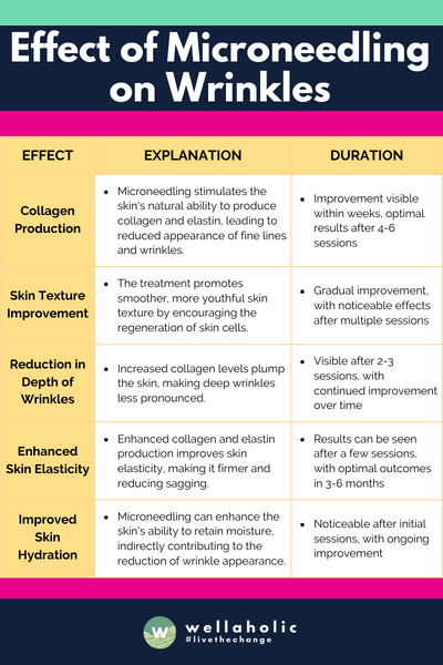
Animal models and in vitro examination of human tissue demonstrate that Microneedling creates micro-channels and micro-wounds at the level of the dermis breaking compact, thickened collagen and inducing the wound healing cascade. Micro-channels cause little epidermal damage making Microneedling safe to use in darker skin phototypes. Gene expression profiling before and after Microneedling treatment demonstrates an upregulation in type I collagen expression, as well as glycosaminoglycans, vascular endothelial growth factor (VEGF), fibroblast growth factor (FGF)-7, epidermal growth factor (EGF), and transforming growth factor (TGF)-β, all important signaling molecules for collagen production, as well as neovascularization. Tissue histology after Microneedling shows thickened epidermis, and an increase in dermal collagen and elastic fiber deposition. Over a period of weeks to months, newly formed type III collagen becomes mature type I collagen causing skin tightening and a decrease in the appearance of scars or rhytides.6–11 In this systematic review, we will explore the efficacy and safety of Microneedling for the treatment of scarring.
Methods
A systematic review using the PRISMA (Preferred Reporting Items for Systemic Review and Meta-Analysis) was completed using the Medline database in August 2020 using the search terms “microneedle” or “microneedling” or “micro needle” or “micro needling” and “scar”. Included manuscripts were case reports, case series and trials discussing the use of microneedling (rolling device, stamping device, fractional radiofrequency device) for the treatment of scars in humans in English. Reviews, animal models, and in vitro studies were excluded, as were articles not available in English.
Summary of Protocols from Studies Discussing the Use of Microneedling for Scar Treatment
| Treatment Protocol | Combination Therapies | |||
|---|---|---|---|---|
| Microneedling (n=36) | Number of passes | 4–10 | Procedural | Platelet-rich plasma (PRP; n=6) Subcision (n=3) Incobotulinum toxin (n=1) Polymethylmethacrylate-collagen gel (n=1) |
| Clinical endpoint | Uniform pinpoint bleeding | Topicals or peels | Glycolic acid peel (n=3) Trichloroacetic acid peel (n=2) Amniotic fluid-derived mesenchymal stem cell, topical (n=1) Hyaluronic acid (n=1) Jessner’s solution peel (n=1) Vitamin A and C, topical (n=1) | |
| Needle depth | 1.5–3.0 mm | Systemic therapy | Systemic isotretinoin (n=1) | |
| 1–12 treatment sessions, every 1–8 weeks | ||||
| Microneedle patch (n=2) | Continuously for 30 days (n=1), changed every 2–3 days for 4–6 weeks (n=1) | |||
| Fractional radiofrequency microneedling (insulated and non-insulated, multiple and single-needle heads, n=20) | Number of passes | 2–8 | Injectable/procedural | Fractional carbon dioxide (CO2; n=2) Fractional 1550 nm erbium-glass (Er:glass; n=1) Hyaluronic acid (n=1) Subcision (n=1) |
| Clinical endpoint | Wheal-like papules/plaques | Topicals | Poly-lactic acid (PLA; n=1) | |
| Needle depth | 0.8–3.5 mm | Systemic therapy | Systemic isotretinoin (n=1) | |
| Energy | 6.82–70 W, 40–82 mJ | |||
| Duration | 50–400 ms | |||
| Total pulses | 250–500 | |||
| 1–6 treatment sessions, every 4–12 weeks |
In all cases of Microneedling or fractional radiofrequency microneedling treatment, patients received pre-treatment anesthesia. Although the most commonly reported anesthesia was topical (lidocaine and/or prilocaine) applied in the area to be treated with or without occlusion for a total of 30 to 90 minutes, 2 studies originating from Germany mentioned the use of general anesthesia. Prior to starting Microneedling, areas were cleansed with either isopropyl or ethyl alcohol. Two studies utilizing fractional radiofrequency microneedling used epidermal cooling to decrease epidermal damage during the procedure. Post-treatment regimens suggested included sunscreen (n=19 studies), bland emollient (n=11), topical or systemic antibiotic including topical fusidic acid (n=11), topical corticosteroids (n=3), cold packs (n=2), as well as topical benzoyl peroxide, cyclopentasiloxane/cyclohexasiloxine/sodium hyaluronate, hyaluronic gel, L ascorbic acid/α-tocopherol/ferulic acid, non-steroid anti-inflammatory drugs (NSAIDs), and tretinoin/kojic acid/hydroquinone/hydrocortisone (n=1 each).6–63
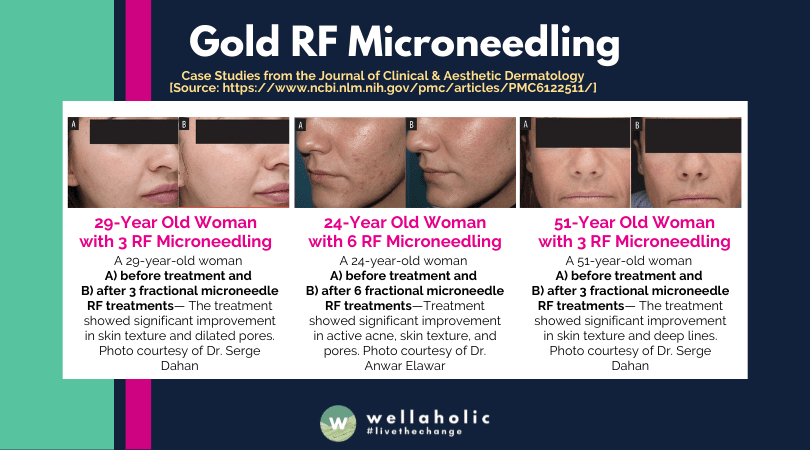
Acne scarring was by far the most discussed condition (n=43 studies). In all studies, boxcar (U-shaped) and rolling (M-shaped) scars demonstrated the greatest clinical improvement after MN or fractional radiofrequency microneedling, while icepick (V-shaped) scars were often recalcitrant.6,9,11–51 Patients treated with MN or fractional radiofrequency microneedling for other types of scars also demonstrated clinical improvement. Scar improvement was measured using both patient and investigator qualitative assessments, as well as the Echelle d’evaluation Clinique des Cicatrices d’Acne (ECCA), Vancouver Scar Scale (VSS), and Visual Analog Scales (VAS).6–63 Combination treatment with laser, PRP, subcision, glycolic acid peel, Jessner’s peel, trichloroacetic acid peel and topical amniotic fluid stem cells resulted in greater scar improvement than Microneedling or fractional radiofrequency microneedlingalone.8,17,24,26,31,32,34,37,40,51,53,55 Fifty to 100% of patients were satisfied with MN or fractional radiofrequency microneedling treatment; 33% of patients reported they would want further treatment,9 while 94% would recommend treatment to others.28 Combining treatment with 1550 nm laser or PRP resulted in higher patient satisfaction.8,34 In addition, patients preferred Microneedling to intralesional triamcinolone (ILTAC) or 1450 nm diode laser.30,56
Adverse events (AEs) due to Microneedling or fractional radiofrequency microneedling were mostly of minimal severity; no serious AEs were reported. Almost all studies reported pain and bleeding during the procedure; the most common post-procedure AEs included transient post-procedure pain/discomfort/burning, erythema and/or swelling. Further AEs are discussed in Table 2. Post-inflammatory hyperpigmentation occurred in 19 studies; 54.5% of studies used FRMN, while 45.5% used Microneedling. There was one case of herpes simplex reactivation which was successfully treated with oral valacyclovir. The most feared AE of Microneedling treatment is the so-called “railroad” or “tramtrack” scarring that can occur with aggressive treatment, and was only reported as an AE in 5 studies.6–63 One female patient with atrophic acne scars developed scarring after an allergic reaction to the nickel contained in the needles, which was subsequently treated with oral prednisolone and topical steroids.38
Summary of Rare AEs Occurring with Microneedling Treatment for Scars
| Adverse Event Description | Number of Studies Reporting (n) |
|---|---|
| Post-inflammatory hyperpigmentation | 19 |
| Scabbing/crusting | 9 |
| Purpura/ecchymosis | 5 |
| “Tramtrack”/“railroad” scarring | 5 |
| Acne flares | 4 |
| Cervical lymphadenopathy | 3 |
| Milia | 2 |
| Pustules/bullae | 2 |
| Allergic reaction to nickel | 1 |
| Herpes simplex reactivation | 1 |
Discussion
Microneedling has gained popularity as a minimally invasive aesthetic technique for the treatment of skin aging, scarring, striae, and hair loss, amongst other indications. Although the US Food and Drug Administration (FDA) initially classified Microneedling as class I medical devices, recent developments have elevated their classification to class II (special controls) and they are currently approved for microdermabrasion, scarring and rhytides.64 The literature suggests that Microneedling and fractional radiofrequency microneedling are well tolerated and result in clinical improvement of scarring due to acne or other infectious cause, hypertrophic or keloid scars, and post-operative or traumatic scars, as well as high rates of patient satisfaction. Microneedling and fractional radiofrequency microneedling were reportedly tolerated better by patients than their laser resurfacing counterparts, namely the CO2, Er:glass and diode lasers, with less reported downtime. Microneedling and FRF-MN can be combined with a variety of other surgical therapies including laser resurfacing, chemical peels, PRP, filler and botulinum toxin for greater clinical results.6–63
As with many aesthetic procedures, Microneedling and fractional radiofrequency microneedling suffer from a lack of standardized protocol. Animal and human studies suggest that multiple passes per treatment and multiple treatment sessions demonstrate greater skin regeneration potential.65,66 Further clinical studies need to be completed to determine optimal number of passes, number of treatment sessions, intertreatment duration intervals and maintenance therapy.
Just like non-ablative laser techniques, Microneedling is an effective intraepidermal and intradermal delivery method for pharmaceuticals.5,67 In addition to microneedles designed to contain substances such as bleomycin or triamcinolone for treatment hypertrophic scarring,68,69 Microneedling can enhance the penetration of topicals such as anesthetics, chemical peels, PRP or filler material such as hyaluronic acid as evidenced by this review,6–63 and possibly nanoparticles and siRNA in the future.67,68 Advances in Microneedling delivery, such as the development of patches or use of MN to deliver energy sources below the level of the epidermis have both decreased the amount of discomfort associated with treatment, and increased efficacy by combining multiple treatment modalities.56,58,70 It is important to note that certain topical products may cause allergy, and even granulomatous reaction, when introduced into the skin through micro-channels created by Microneedling devices; physicians should counsel patients appropriately regarding these risks. Substances such as bleomycin, triamcinolone and filler material should presumably be safer to administer through micro-channels given that they are designed as injectables.
Although no serious AEs were associated with Microneedling and fractional radiofrequency microneedling treatment of scars, it is important to note that post-inflammatory hyperpigmentation occurred in over 30% of studies and resolved either spontaneously or with the help of topical bleaching creams within months. Given that the majority of patients treated in this review had a Fitzpatrick skin phototype of IV or greater, AE reporting may have been biased towards events that more commonly occur in this patient groups post-procedure, specifically dyspigmentation and aberrant scarring (such as the “tramtracks”). Physicians should be aware that Microneedling is not without its risks, and appropriately counsel patients during the consent process to avoid patient morbidity post-treatment.
Limitations of this study include its lack of meta-analysis. Given the heterogeneity of data presented by included studies, it was not possible to combine and statistically analyze this data.
Conclusion
Microneedling and its relative fractional radiofrequency microneedling are both well-tolerated, minimally invasive procedures that can be used for the effective treatment of scarring. Although there are no standard treatment protocols, clinical improvement in many types of scars including acne, varicella and smallpox, hypertrophic or keloid, and post-operative or post-traumatic scars have been reported in the literature.
Microneedling and fractional radiofrequency microneedling can be used as stand-alone modalities, or can be combined with a variety of topicals and other surgical procedures for superior results. No serious AEs have been reported using Microneedling or fractional radiofrequency microneedling for the treatment of scars; however, physicians should be aware that post-inflammatory hyperpigmentation is still a relatively common event. Large-scale, clinical trials need to be completed to determine optimal, standardized protocols to treat scarring using Microneedling or fractional radiofrequency microneedling.
Source
- National Library of Medicine | Clin Cosmet Investig Dermatol. 2020; 13: 997–1003.
- Microneedling for the Treatment of Scars: An Update for Clinicians
- Margit L W Juhasz1 and Joel L Cohen1,2

Serene Chiam, Aesthetic Director
Serene Chiam is the Aesthetic Director at Wellaholic, a well-known aesthetic chain in Singapore. She has more than ten years of experience in the aesthetics industry. With a Bachelor of Health Science (Aesthetics) and CIDESCO certifications, she expertly combines scientific knowledge with practical skills. Serene is known for her personalized approach to beauty, ensuring each Wellaholic client’s journey is unique and transformative. Her significant contributions have been pivotal in establishing Wellaholic’s reputation for excellence in aesthetic wellness.
Contact Serene at support@wellaholic.com
GET IN TOUCH
Book Now Pay Later

Gold RF Microneedling Facial
- ⭐ Uses Up to 64 Micro Needles. Gold RF Microneedling: Ultimate anti-aging treatment with 64 needles to penetrate the skin, release RF energy, and trigger collagen and elastin production for a clearer complexion and firmer skin
- ⭐ Safe and Minimally Invasive. Gold RF Microneedling is a safe, minimally invasive.
- ⭐ Effectively Treats Acne Scars, Pigmentation & Wrinkles. Extremely effective aesthetic treatment for treating acne scars, pigmentation, fine lines and wrinkles.
- ⭐ Stimulates Collagen Growth. Gold RF Microneedling stimulates collagen and elastin for new, youthful-looking skin.
- ⭐ Award-Winning. Wellaholic’s treatments have been recognized by top beauty publications such as Daily Vanity, Beauty Insider, and Tropika Club Magazine.
- ⭐ Over 2000 Verified Customer Reviews. Wellaholic has over 30 industry awards and over 2000 positive reviews from customers, and >50% are repeat customers.